
Retroviral Pathogenesis: HTLV-1 and Adult T-Cell Leukemia
More details: https://www.thoma-kress-lab.de/
Human T-cell leukemia virus Type 1 (HTLV-1)
Our group studies HTLV-1, a highly oncogenic yet neglected retrovirus, which primarily infects white blood cells (CD4+ T-cells) in vivo and causes incurable diseases like Adult T-cell leukemia/lymphoma (ATLL) or inflammatory maladies like HTLV-1-associated myelopathy/tropical spastic paraparesis (HAM/TSP) after lifelong viral persistence. Worldwide, at least 5-10 million people are HTLV-1 infected and most of them are unaware of their infection. Endemic regions are located in Japan, Central Australia, Melanesia, South America, the Caribbean, and the Middle East. Virus prevalence is low in Germany (Korn et al., 2026). The virus is transmitted via cell-containing body fluids such as blood products, semen and breast-milk, which constitutes the major route of mother-to-child transmission. After infection of CD4+ T-cells, HTLV-1 is reversely transcribed and integrates into the host cell genome. Despite the high number of people worldwide living with a persistent HTLV-1 infection, there is still no efficient prevention strategy, vaccine or cure for HTLV-1 and its associated diseases.
Our major research aims are:
- Understand breast milk transmission of HTLV-1 and develop prevention strategies
- Elucidate molecular mechanisms of HTLV-1 cell-to-cell transmission
- Develop strategies to lower the proviral load of infected individuals
- Interfere with HTLV-1 induced leukemogenesis
Milk transmission of HTLV-1 and develop prevention strategies
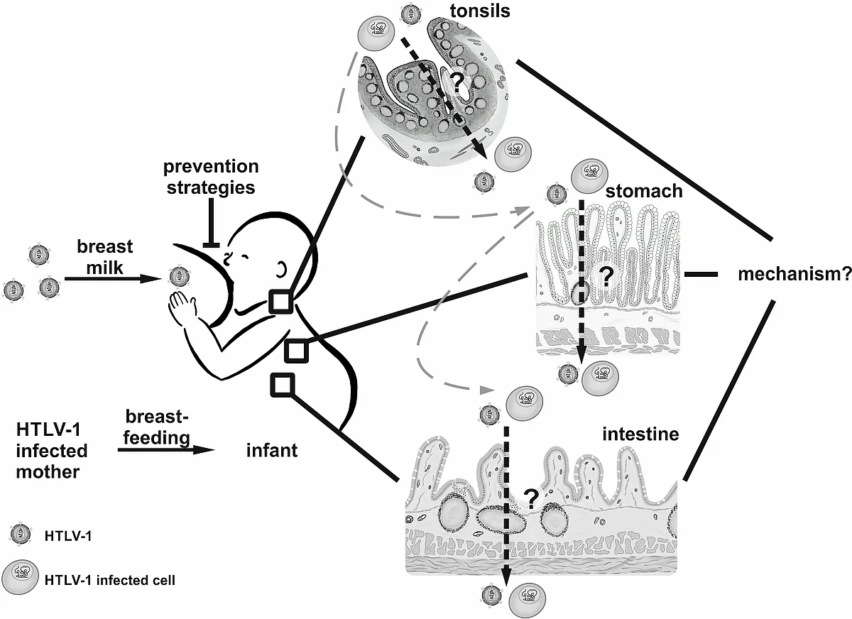
In 2025, we could successfully prolongate our Junior Research Group in Infection Research, which is generously funded by the Federal Ministry of Research, Technology and Space (BMFTR). Within the project Milk-TV: MilkTransmission of Viruses, we seek to explore early events of HTLV-1 transmission from mother-to-child and to develop prevention strategies to fight HTLV-1 infections. Briefly, breastfeeding is recommended by the World Health Organization for at least 6 months and up to 2 years of age, and it is proven that breast milk protects against several diseases and viral infections. Intriguingly, few viruses are preferentially transmitted via breastfeeding including HTLV-1, HCMV and HIV. Risk of transmission increases with the duration of breastfeeding, however, abstinence from breastfeeding is not an option in resource-limited settings or underrepresented areas or populations. Despite significant progress in understanding details of cell-to-cell transmission, it is still unclear, which cells in which organs get infected via the oral route of virus transmission (Kemeter et al., 2023), how these cells get infected, how breast milk affects this route of infection, and how to inhibit oral transmission despite breastfeeding (Figure 1), which is an urgent need, especially in underrepresented areas of the world (Millen and Thoma-Kress, 2022). In the first funding period, we developed different cell and tissue culture model systems in the lab to address these questions (Heym et al., 2024). Specifically, we characterized the impact of milk on cell-to-cell transmission in T-cells and also studied how acidification impacts HTLV-1-infected cells and the so-called viral biofilm, a specialized structure required for HTLV-1 transmission. We have also moved forward in studying virus transmission across epithelial barriers and have started to investigate viral dissemination in different organs of the gastrointestinal tract in vivo. Finally, we have developed HTLV-1 specific nanobodies in collaboration with Dr. I. Fernandez (Institute Pasteur, Paris), which we optimize in their biochemical properties in the current funding period of the project.
Elucidate molecular mechanisms of HTLV-1 cell-to-cell transmission
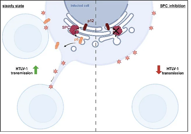
Transmission of HTLV-1 to other T-cells is strictly dependent on cell-cell contacts, and viral particles are transferred after polarized budding at a tight and confined cell-cell contacts, the so-called virological synapse or via viral biofilm formation. However, HTLV-1 is not only transmitted at tight cell-cell contacts, rather, transmission may also occur via long-distance connections, which are induced by the small accessory viral protein p8. This protein is generated by proteolytic cleavage from the precursor p12 encoded on open reading frame I of the HTLV-1 genome. p8 induces cellular protrusions and is transferred to other cells to foster HTLV-1 cell-to-cell transmission. Interestingly, many viruses exploit cell-cell communication pathways to transfer viral proteins to other cells independent of viral particle formation (Simon and Thoma-Kress, 2024). We could previously identify novel interaction partners of p8 including vasodilator stimulated phosphoprotein (VASP) and other related proteins, which were important for p8 transfer between cells and for virus transmission (Donhauser et al., 2020).
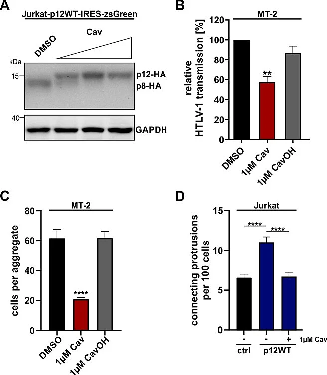
In our recent work, we have identified how p8 is cleaved from its precursor p12 (Simon et al. 2025; Figure 2). Combining bioinformatics with wet-lab work, we could show that p12 contains a signal peptide cleaved by the host Signal Peptidase Complex (SPC) to produce p8. SPC inhibition with Cavinafungin, SPC knockdown, and signal-peptide mutations prevented p12 cleavage, reduced cell aggregation and conduit formation, and impaired viral transmission from MT-2 cells to Jurkat T cells. These results identified the SPC as the host factor generating p8 and demonstrated that blocking p12 cleavage disrupts p8-dependent HTLV-1 cell-to-cell transmission (Figure 3).
Importantly, based on these novel findings, we were able to generate the first p12-only mutants, i.e. cleavage deficient p12 mutants. These mutants are now a unique tool to study the role of p12 and p8 independently of each other.
Develop strategies to lower the proviral load of infected individuals
Our lab works on different projects to interfere with HTLV-1 persistence to ultimately lower the proviral load. This is of special importance since the risk to develop HTLV-1 associated diseases increases with higher proviral loads. Upon infection with HTLV-1, the viral transactivator Tax stimulates viral replication by recruiting host cell factors like positive transcription elongation factor b (p-TEFb) to the viral promoter and by enhancing mitotic expansion of infected cells. Since viral gene expression is repressed in vivo by viral, cellular, and epigenetic mechanisms in late phases of infection, HTLV-1 avoids an efficient CD8+ cytotoxic T-cell (CTL) response directed against the immunodominant viral Tax antigen. Hence, new therapeutic strategies aim to transiently activate viral gene expression to shift the equilibrium in favor of an enhanced CTL response to enhance immunogenicity of HTLV-1 Tax, and thus, to expose the latent HTLV-1 reservoir to immune destruction. Thus far, the composition of the protein complex guiding viral gene expression is only partially understood, and systematic analyses and comparisons of compounds affecting viral transcription are lacking. In earlier work, we could identify strong and specific upregulation of the transcription elongation factor ELL2 in HTLV-1-infected cells (Mann et al., 2014). We found that ELL2 strongly enhances Tax-mediated transactivation of the HTLV-1 promoter, and that ELL2 and Tax are part of a common protein complex. We uncovered important domains within Tax and ELL2, which are crucial for Tax:ELL2 complex formation (Kohrt et al., 2021). Moreover, we analyzed the subcellular localization of ELL2 in more detail and could identify a very strong nuclear localization signal (NLS) in ELL2. Introducing single point mutations into this NLS sequence completely changed the subcellular localization of ELL2 and led to its distribution in the cytoplasm (Kohrt et al., 2024). Based on these findings, we analyzed the interplay between Tax and ELL2 on transcript and protein level in more detail and found that both proteins influence each other with regard to subcellular localization. To develop strategies to interfere with HTLV-1 transcription and to enhance immunogenicity of HTLV-1, we analyzed histone deacetylase inhibitors and identified Panobinostat and Romidepsin to be superior to previously tested compounds in enhancing viral transcription (Schnell et al., 2022). In another project, we are currently assessing alternative strategies to interfere with HTLV-1 transcription. In a collaborative project, we attacked the integrated HTLV-1 provirus by molecular evolution of a designer recombinase, which was adapted to a recognition site within the HTLV-1 long terminal repeats flanking the integrated proviral DNA (Rojo-Romanos et al., 2023).
Interfere with HTLV-1-induced leukemogenesis
It has been known for years that constitutive activation of the classical and alternative NF-κB signaling pathways by the viral oncoprotein Tax is a hallmark of HTLV-1-driven cancer. NF-κB-deficient Tax transgenic mice lack the induction of Adult T-cell leukemia/lymphoma (ATLL)-associated aggressive skin diseases. Further, animal studies therapeutically targeting NF-κB slow down and reduce tumor growth in ATLL-like diseases. Although there are different reports whether NF-κB is critical for initiating cellular transformation, there is a strong connection between Tax, NF-κB, tumor formation and maintenance. We could show that activation of NF-κB signaling specifically enhances the abundance of Tax protein, but not of Tax transcripts (Millen et al., 2020). This led to the identification of a positive feedback loop between Tax and NF-κB activity, which results in enhanced protein expression of Tax and might thus serve as a novel therapeutic target to interfere with Tax-driven transformation. In a collaboration with Dr. Calabro (Padova, Italy), we analyzed the interplay between HTLV-1-infected T-cells and the tumor microenvironment making use of a mouse model of ATLL, and analyzed the impact of the actin-bundling protein Fascin on HTLV-1-induced leukemogenesis. Fascin is NF-κB-dependently induced by Tax (Heym et al., 2022) and contributes to invasive migration of HTLV-1-infected cells (Kress et al., 2011). In future projects, we seek to analyze the role of Fascin in leukemogenesis in context of the human immune system.
AG Thoma-Kress in the media:
- ImmunoTeaTime. 18.08.2025. Podcast with Andrea: Milk TV: Fighting HTLV ft. Dr. Andrea Thoma-Kreß Meilenstein im Kampf gegen Auslöser einer Form der Leukämie. 28.03.2023.
https://www.virologie.uk-erlangen.de/aktuelles/nachrichten/detail/meilenstein-im-kampf-gegen-ausloeser-einer-form-der-leukaemie/ - Neue FAU-Nachwuchsgruppenleiterin: Dr. Andrea Thoma-Kreß. 30.3.2021.
https://blogs.fau.de/gsfau/2021/03/neue-nachwuchsgruppenleiterin-dr-andrea-thoma-kress/ - Unsere FAU-Nachwuchsgruppenleitungen. 30.3.2021
Unsere FAU-Nachwuchsgruppenleitungen | Friedrich-Alexander-Universität Erlangen-Nürnberg - Wege in die Wissenschaft. 24.2.2021. FAU-Magazin “Friedrich”.
https://www.fau.de/2021/02/friedrich/wege-in-die-wissenschaft/ - Virus im Fokus. Infektionsforschungsprojekt profitiert von Nachwuchsgruppenförderung durch das BMBF. 16.11.2021.
https://www.fau.de/2020/11/news/wissenschaft/virus-im-fokus/ - Der Weg ist das Ziel. Ampuls 3/2020, Seite 7. Zeitung für alle Mitarbeitenden des Uni-Klinikums Erlangen.
https://mitarbeiterportal.intranet.uk-erlangen.de/display/AKTUELLESpub/Archiv?preview=/5715115/158270003/ampuls_2020_3.pdf - Auszeichnung für herausragende Grundlagenforschung zur Tumorentstehung. 5.10.2017.
https://www.fau.de/2017/10/news/panorama/auszeichnung-fuer-herausragende-grundlagenforschung-zur-tumorentstehung/ - LinkedIn: Andrea Thoma-Kress
Andrea Thoma-Kress | LinkedIn