Molecular Mechanisms of Herpesvirus Entry and Fusion
Our group moved from the German Primate Center (Göttingen) to the Harald zur Hausen Institute of Virology in 2025. Our previous and ongoing research focuses on the early steps of viral infection. We investigate the role of viral glycoproteins and their cellular receptors in attachment, entry, and membrane fusion. Our overarching goal is to understand how these interactions determine cell and tissue tropism and how they may serve as targets for neutralizing antibodies.
Kaposi’s sarcoma-associated herpesvirus (KSHV)
KSHV is an oncogenic human γ-herpesvirus and the etiological agent of Kaposi’s sarcoma, primary effusion lymphoma, and a plasmablastic variant of multicentric Castleman’s disease. KSHV infection is highly prevalent in Sub-Saharan Africa and represents a major global health burden.
A detailed mechanistic understanding of how the different KSHV envelope glycoproteins and their receptors mediate viral attachment, entry, and membrane fusion is essential not only for understanding viral biology but also for the rational design of vaccines and antiviral agents such as neutralizing antibodies.
In addition to KSHV, we study the closely related rhesus monkey rhadinovirus (RRV), which serves as a valuable comparative model. RRV allows informative side-by-side analyses to distinguish conserved from virus-specific features of entry and fusion.
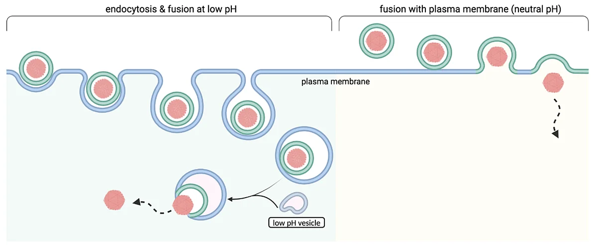
Enveloped viruses can fuse either after endocytosis or directly at the plasma membrane. (from Liu et al., PLoS Pathog., 2025, created using biorender.com)
Function and regulation of the herpesviral core fusion machinery
Herpesviruses are enveloped viruses surrounded by a lipid membrane in which viral glycoproteins are embedded. These glycoproteins bind to cellular receptors that mediate attachment, trigger endocytosis, or initiate membrane fusion – the final step required to release the capsid into the cytoplasm.
The conserved herpesviral core fusion machinery consists of glycoproteins gB, gH, and gL. Its regulation is complex and incompletely understood. gB acts as the fusion protein and catalyzes membrane merger by transitioning from a metastable pre-fusion conformation to a stable post-fusion conformation.
KSHV adds an additional layer of complexity: according to our recent work (Liu et al., 2025), KSHV requires low pH as a fusion trigger and does not efficiently induce cell–cell fusion at neutral pH.
Using RRV for comparative studies, we demonstrated that RRV can utilize both direct plasma membrane fusion and low-pH-triggered fusion after endocytosis, and it exhibits robust cell–cell fusion activity that is further enhanced under acidic conditions. By swapping gB between the two viruses, we showed that KSHV gB dictates the strict dependency on low pH that is observed for KSHV infection.
Ongoing structure–function studies aim to identify the molecular features that restrict KSHV entry to a low-pH endocytotic pathway. In parallel, we are working to stabilize gB in its normally labile pre-fusion conformation and to analyze the functional and antigenic differences between its conformational states.
Receptor tropism and cell-type specific entry requirements
We identified Tim-1 and Tim-4 as potent receptors for KSHV and the related RRV. In addition, we performed animal studies demonstrating a tissue-specific role for the KSHV glycoprotein K8.1 and establishing the suitability of the common marmoset model to study tissue tropism as well as cellular and humoral immune responses to KSHV. These studies are currently ongoing, preliminary results are already available as preprints (https://www.biorxiv.org/content/10.1101/2023.10.10.561662v2, https://www.biorxiv.org/content/10.1101/2024.10.31.621273v1).
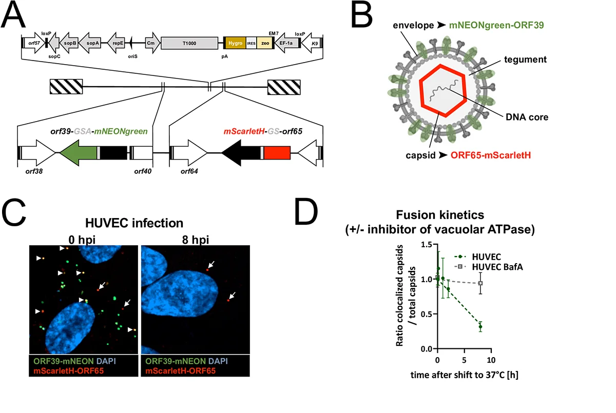
A) Map of BAC 16 noGFP_mScarletH-ORF65_ORF39-mNeonGreen encoding KSHVmS65-39mN (not exactly to scale). B) Schematic representation of dual-labelled virions (not to scale). C) HUVEC cells imaged after virus attachment in the cold 0 hours post infection (hpi) or after incubation at 37°C at 8 hpi. Arrow heads indicate colocalization of mScarletH-ORF65 and ORF39-mNeonGreen, arrows indicate single positive capsids. hpi: hours post infection. D) Virus fusion with HUVEC was analysed by calculating the ratio of envelope-colocalized capsids to total capsid in the absence or presence of Bafilomycin A1 (BafA), an inhibitor of the vacuolar ATPase. (from Liu et al., PLoS Pathog., 2025)
Molecular tools for studying entry and fusion
To visualize attachment, endocytotic uptake, and membrane fusion we developed mono- and dual-fluorescent KSHV reporter viruses (Liu et al., 2025).
By tagging the capsid with a red fluorescent protein and an envelope protein with a green fluorescent protein, loss of colocalization can be used as a directly observable surrogate marker for membrane fusion. Upon membrane fusion, the fluorescently tagged envelope proteins rapidly diffuse within the cellular membrane, whereas the capsid continues intracellular trafficking toward the nucleus.